

The American College of Radiology (ACR) gave its recommendations on managing splenic incidentalomas on CT or MRI in 2013. These can be accessed (paid access) by clicking on the link here. The guidelines are, in my humble opinion, a bit conservative, but nevertheless this is what we have to follow as of now.
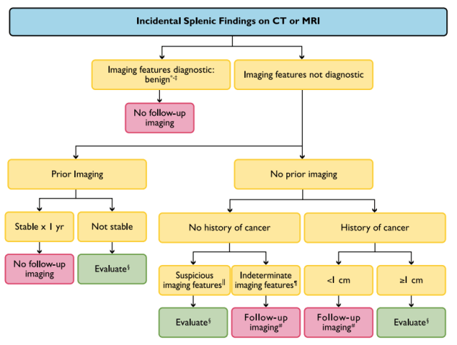
*Cyst: imperceptible wall, near-water attenuation (10 HU), no enhancement.
†Hemangioma: discontinuous, peripheral, centripetal enhancement (findings that are uncommon in splenic hemangiomas).
‡Benign imaging features: homogeneous, low attenuation (20 HU), no enhancement, smooth margins.
§Evaluate: PET vs. MRI vs. biopsy. Suspicious imaging features: heterogeneous,
enhancement, irregular margins, necrosis, splenic parenchymal or vascular invasion, substantial enlargement.
¶Indeterminate imaging features: heterogeneous, intermediate attenuation (20 HU), enhancement, smooth margins.
#Follow-up MRI in 6 and 12 months
– Akshay Baheti, Tata Memorial Hospital
PS: All images are a copyright of the original published article.

Pingback: Incidental Findings Follow-up Recommendations – Cafe Roentgen