

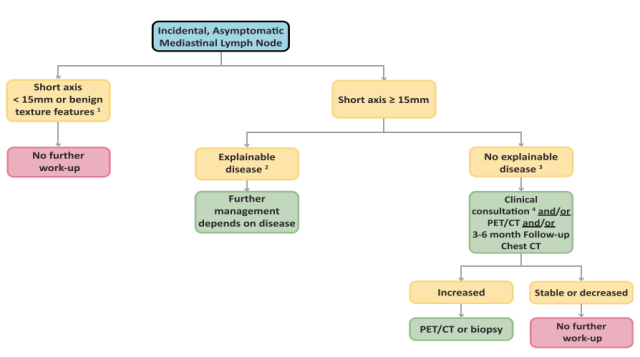
The American College of Radiology (ACR) gave its recommendations on managing abdominal nodes on CT or MRI in 2013. These can be accessed (paid access) by clicking on the link here. The management algorithm for mediastinal nodes has also been subsequently published in 2018 (open-access), which you can read here in more detail. The important point to remember is that the short axis cut-off is 10 mm for retroperitoneal nodes while it is 15 mm for mediastinal nodes.
You can read our blog on the ACR guidelines for incidental mediastinal findings (including masses and pericardial abnormalities) here.
Management of incidental abdominal lymph nodes
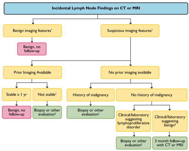
*Benign imaging features: normal short-axis diameter (1 cm in retroperitoneum), normal architecture (elongated, fatty hilum), normal enhancement, normal node number.
†Abnormal imaging features: enlarged short-axis diameter (1 cm in retroperitoneum), architecture (round, indistinct hilum), enhancement (necrosis/hypervascular), increased number (cluster of 3 lymph nodes in single nodal station or cluster of 2 lymph nodes in 2 regions).
‡Nonneoplastic disease: eg, infection, inflammation, connective tissue.
§Other evaluation (PET/CT, nuclear scintigraphy [MIBG], endoscopic ultrasound).
Evaluation of Incidental Mediastinal Node
– Akshay Baheti, Tata Memorial Hospital
PS: All images are a copyright of the original published article.

Pingback: Incidental Findings Follow-up Recommendations – Cafe Roentgen