

The ACR published its recommendations for managing incidental hepatic lesions seen on CT in 2017. The salient feature of the guidelines is that subcm sized incidental liver lesions in patients without any risk factors need no further evaluation. The overall guidelines are given below; the original article is open-access and available here for further reading.
A word of caution: The US doesn’t use ultrasound as it should, and you will find the guidelines very MRI heavy. In the context of ultrasound friendly regions like India or Europe, the guidelines need not be adhered to rigidly, and the use of ultrasound must be optimized. This, of course, is a personal opinion.
Incidental Liver Lesion <1 cm

Incidental Liver Lesion 1-1.5 cm

Incidental Liver Lesion >1.5 cm

Note:
Patient risk categorization
| Low-risk patients
|
No known malignancy
No hepatic dysfunction No hepatic risk factors |
| High-risk patients
|
Known malignancy with a propensity to metastasize to the liver
Cirrhosis Presence of hepatic risk factors* |
* Hepatic risk factors: hepatitis, nonalcoholic steatohepatitis, alcoholism, sclerosing cholangitis, primary biliary cirrhosis, choledochal cysts, hemochromatosis and other hereditary hepatic conditions, and anabolic steroid use
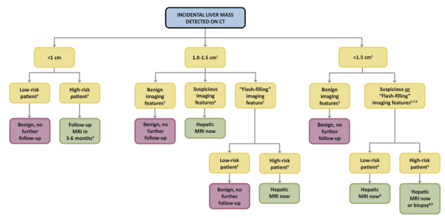
Benign features: sharp margins, homogeneous low density (<20 HU) on noncontrast and/or portal venous–phase, and characteristic features of hemangiomas, FNH, focal fatty sparing or deposition, or perfusional changes.
Suspicious features: ill-defined margins, heterogeneous density, mural thickening or nodularity, thick septa, and intermediate to high attenuation on portal venous–phase imaging (>20 HU, in the absence of pseudoenhancement). If pre- and post contrast CT is available, enhancement >20 HU is a suspicious feature. To evaluate, prefer MRI.
“Flash-filling” feature: uniform hyperenhancement relative to hepatic parenchyma on arterial-phase (including late arterial/early portal venous–phase) imaging. If additional postcontrast phases are available to characterize lesion as benign (eg, hemangioma) or suspicious (eg, HCC), the lesion should be placed in one of those respective categories.
Overall ACR Algorithm for Incidental Liver Lesions
– Akshay Baheti, Tata Memorial Hospital
PS: All images are a copyright of the original published article.

Pingback: Incidental Findings Follow-up Recommendations – Cafe Roentgen
Hi friends, its impressive post concerning teachingand entirely explained,
keep it up all the time.
LikeLike