

Based on the talk by Dr Aditya Daftary and Dr Malini Lawande, here is a checklist to comprehensively describe a fracture.
Checklist to describe fractures:
- Site:
-
- Epiphysis, metaphysis, diaphysis
- Proximal/mid/distal shaft
- Orientation:
-
- Transverse/oblique/vertical (avoid “comminuted” unless it is truly completely fragmented)
- Angulation
-
- Apex medial/lateral/anterior/posterior
- Varus/valgus
- Displacement of distal fragment and extent
-
- Minimal/How many shaft width (1/2, full)
- Articular extension, gap and step off
- Associated soft tissue swelling
For example, the following fractures would be described as follows:

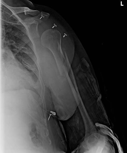
Frontal radiograph of the left shoulder. Non-displaced left greater tuberosity fracture. No intra-articular extension.
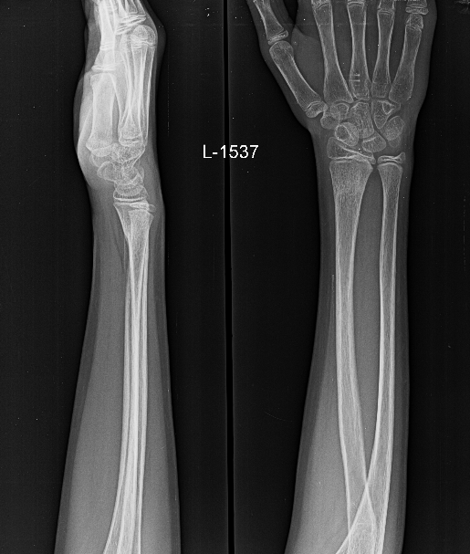
Frontal and lateral radiographs of the left forearm and wrist in an immature skeleton. Subtle buckling of the left distal radius lateral cortex without extension to the medial cortex or angulation, in keeping with torus fracture. There is associated anterior bowing of the pronator quadratus fat pad, consistent with deep soft tissue edema.
(NOTE: Read more on the pronator quadratus sign at https://radiopaedia.org/articles/pronator-quadratus-sign)
Frontal radiograph of the left humerus . Obliquely oriented proximal left humeral shaft fracture with half shaft width lateral displacement.
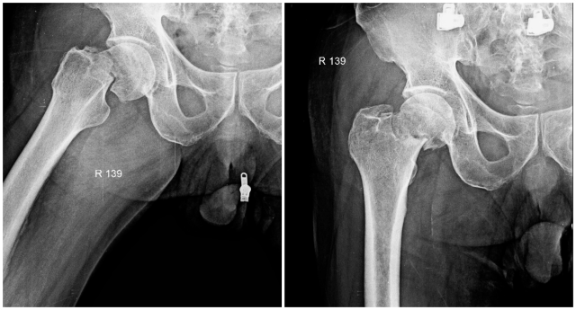
Frontal and frog leg lateral radiographs of the right hip. Transversely oriented right femoral neck fracture with one third shaft width lateral displacement and no intraarticular extension.
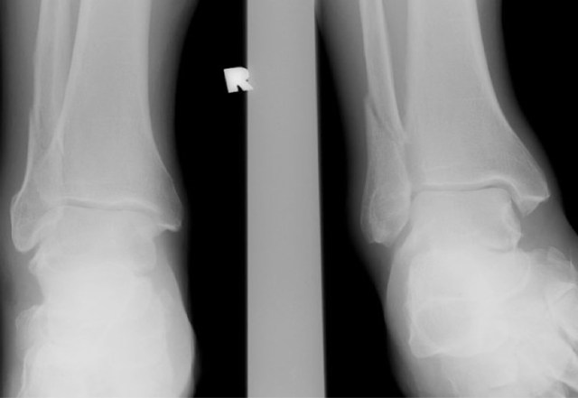
Frontal and mortise radiographs of right ankle. Essentially non-displaced right suprasyndesmotic distal fibular shaft fracture. No overt lateral or medial ankle joint space widening.

Pingback: The Art and Science of the Radiology Report – Cafe Roentgen