
Do you like cheese? Parmesan, mozzarella, cream, cottage, cheddar – the mouth starts watering at the very mention of cheese – a dairy product whose origins predate recorded history. From pizza toppings to sandwich layers and indulgent dishes like shahi paneer, cheese holds a cherished place in global cuisine. But did you know that there is one variety of cheese called camembert which played a vital role in the development of ultrasound elastography? Yes, you read that right. Apart from gracing our plates, cheese has also revolutionised how we assess liver fibrosis, lymph nodes, breast, thyroid and prostate nodules, among so many other things. This little-known story begins in France.
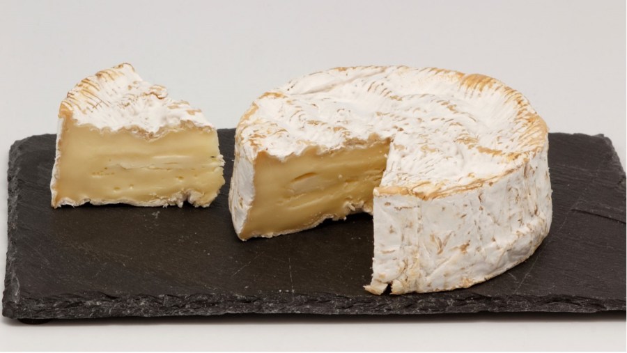
Originating from the beautiful countryside of Normandy, camembert is a soft, creamy variety of cheese with a velvety texture and an earthy flavour profile. It is made from cow’s milk and undergoes a rigorous aging process, during which it develops its characteristic solid rind and a gooey interior. Traditionally, this cheese was made from raw, unpasteurised milk of Normandes cows; however, due to health concerns, most varieties outside Europe, especially in the United States, are now prepared from pasteurised milk.
The credit for the invention of the camembert goes to Marie Harel, a skilled cheesemaker from Normandy, who refined a cheese recipe shared with her by Abbot Charles-Jean Bonvoust, a former priest with a passion for cheesemaking. Marie’s descendants continued her legacy, producing camembert on a larger scale. The cheese gained regal approval when her son-in-law presented a sample to Napoleon III, who endorsed it with his royal seal.
In recognition of her contribution, Marie Harel was commemorated with a Google Doodle on her 256th birthday in 2017. Camembert’s buttery, savoury taste goes flawlessly with bread, fruits, or a glass of wine, making it a favourite among both connoisseurs and the general public alike.

Camembert cheese is best enjoyed when it has matured into having a thin external crust and a soft, gooey inside. This balance is key. For cheesemakers, knowing when it’s just right is crucial. For generations, cheesemakers relied on the tactile wisdom of thumb-pressing to assess camembert’s ripeness. However, with the advent of modern technology, a new chapter in cheese evaluation began.

In the late 1990s, French physicist Dr Mathias Fink embarked on a pioneering venture. His goal was simple: to combine shear waves (low-frequency waves with a pushing force) with ultrasound technology to gauge the stiffness and maturity of camembert cheese. With the help of his students, he successfully developed a cutting-edge digital ultra-fast ultrasound imaging system that could generate shear waves at supersonic frequencies and simultaneously measure them. This allowed for an accurate assessment of the elasticity of a sample of camembert cheese in real time.
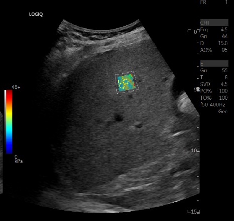
In an interesting turn of events, Dr Fink, alongside his student Mickael Tanter, recognised the potential of this technology in the realm of medical diagnosis. Their ingenuity led to them developing and patenting a technique called ‘shear wave elastography.’ This technique used shear waves to identify subtle differences in the elasticity of human tissues and displayed them in the form of real-time colour-coded maps. These maps offered a non-invasive means to identify diseased tissues within the body, a truly transformative advancement in medical imaging.
In 2005, Fink and Tanter, along with six other co-founders, established a medical imaging start-up called SuperSonic Imagine. After securing their initial round of funding, they developed ten machines, and deployed them in several breast cancer treatment centres for testing. The results were astonishing, to say the least – these machines significantly reduced the number of unnecessary breast cancer biopsies. This technology clearly had the potential to change the landscape of diagnostic procedures.
Encouraged by their initial success, in 2009, they introduced their innovative SWE imaging device to the market. Dubbed “AixPlorer” after the company’s headquarters in Aix-en-Provence, France, this device quickly became a game-changer in the medical field, with a particularly pronounced impact in the evaluation of liver fibrosis.
The rest, as they say, is history. Over the years, ultrasound elastography has dramatically transformed how we assess a variety of pathologies, offering a non-invasive, more comfortable alternative to traditional invasive procedures.
Today, ultrasound elastography is the cornerstone of assessment of liver fibrosis, lymph nodes, cancers of the thyroid, breast and prostate, as well as in musculoskeletal imaging in the evaluation of muscles and tendons. So, the next time someone asks you to do a ‘FibroScan’, smile and think about how it all started – with an endeavour to measure the softness of cheese!
– Dr Anmol Dhawan
Dr Anmol Dhawan is a radiologist working as a senior resident at Bharati Hospital, Pune. He enjoys writing about radiology, history and culture. You can check out his blog at https://syndrome.home.blog/ and you can reach out to him at anmoldhawan@gmail.com.

Fascinating tale.And very well written as well Dr.Dhawan!
LikeLike
Thank you so much. I’m glad you enjoyed it!
LikeLike
Fascinating facts so well expressed! Want to read more of them in future too!
LikeLike
Thank you for reminding us of the beginnings of our adventure in Ultrasound Elastography. We were interested not only in cheese but also in “foie gras”, two very French specialities !!!!!.
Mathias Fink
LikeLiked by 1 person
Had heard that the “FibroScan” was invented for measuring the stiffness of cheese. Now this is a fascinating story! Well written
LikeLike